Nuclear Scintigraphy
Nuclear scintigraphy has also been referred to as the bone scan, nuclear scan, and nuclear medicine. This diagnostic modality is one of the most sensitive tools available for evaluating the musculoskeletal system of the horse. Nuclear scintigraphy is a very sensitive diagnostic tool, allowing us to evaluate both soft tissue and bony structures. There are different options available for nuclear scintigraphy, based on which areas of the horse are of interest. Following is a list of options that the Ontario Equine Hospital offers.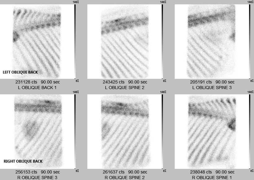
Kissing Spine Lesion
When Should Nuclear Scintigraphy Be Considered?
- Horses with a history of poor performance and no evident lameness
- Horses with a history of lameness or poor performance that is suspected to be multifocal in origin
- When lameness originating from the upper limbs or pelvis is suspected
- Subtle or intermittent lameness
- Lameness that has not been able to be identified or localized with other diagnostics (nerve blocks, radiographs, ultrasound, etc.)
- To monitor the progress of previously diagnosed fractures or other injuries of bone or soft tissue
- Pre-purchase examinations
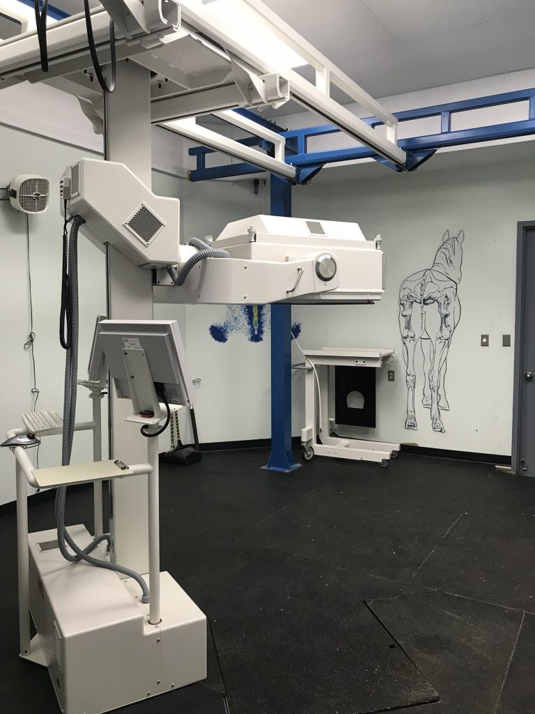
The Nuclear Scan Procedure
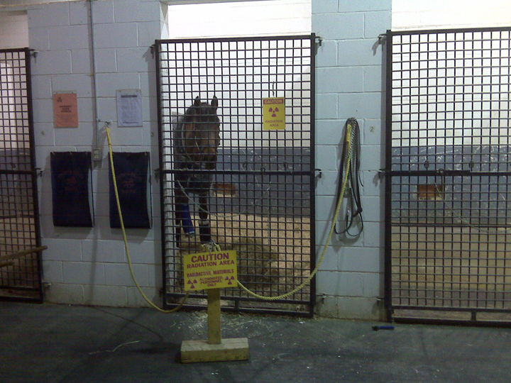
All patients booked for nuclear scintigraphy arrive to the hospital the day before the procedure. This allows us to have an adequate amount of time to prepare the horse for the procedure the following day. All nuclear scans are carried out by either a veterinarian or a Registered Veterinary Technician. Two hours prior to the imaging procedure, the horse is injected intravenously with a radioactive isotope called Technitium (TC-99). This isotope concentrates in areas where there is high metabolic activity of the tissues or inflammation present, also referred to as a “hot spot”. Once the horse is ready to be imaged, sedation is administered to facilitate the process and therefore enable us to maximize the quality of the images obtained.
Nuclear scintigraphy images of the horse are acquired using a Gamma Camera, and areas of abnormal isotope uptake are identified. Areas of abnormal uptake may include chip fractures, stress fractures or remodelling, osteoarthritis (degenerative joint disease), and soft tissue injuries to ligament or tendon structures.